TL;DR ✨
This literature review is only a very short extract from what occupied the author over the preceding few weeks. He attempted to summarize the existing knowledge of keratin and the possible use of its hydrolysate to enrich feed biomass for farmed animals.
Keratin is a mechanically resistant and chemically unreactive macromolecular protein found in all mammals. It is insoluble in water, weak acids, and weak bases, and also dissolves poorly in polar organic solvents.
This literature review is only a very short extract from what occupied the author over the preceding few weeks. He attempted to summarize the existing knowledge of keratin and the possible use of its hydrolysate to enrich feed biomass for farmed animals. While searching for information, he found a significant lack of comprehensive material about keratin on the Czech internet and therefore tried to fill that gap at least partially. The text is an extract from several publications and takes the form of a literature review. The author believes it may occasionally prove useful to someone, which is why he has kept it on his blog. When he has nothing else to do one day, he will add it to the Czech Wikipedia. :-)
1. Description of Keratin
Keratin is a mechanically resistant and chemically unreactive macromolecular protein found in all mammals. It is insoluble in water, weak acids, and weak bases, and also dissolves poorly in polar organic solvents.
Keratin is highly resistant to enzymatic degradation, most commonly by pepsin and trypsin. Degradation is prevented by keratin helices intertwined through disulfide bonds between cysteines, as well as by hydrogen bonds connecting the individual keratin fibers.
Keratin is a basic structural material in all higher vertebrates and belongs to the fibrillar proteins and scleroproteins. It therefore most often serves protective and mechanical functions and occurs mainly in places subject to greater stress where reinforcement is needed.
It is frequently found in epithelial tissue and in cells, where it forms part of the intermediate filaments. These are among the most firmly arranged fibers in organisms. Keratin microfilaments form the basis of microtubules, strong fibrous structures in the cytoskeleton that provide transport for organelles within the cell.
In higher animals it is found, for example, in horns, nails, body hair, wool, and human hair. In reptiles, birds, amphibians, and other animals, keratin occurs in the outer layer covering the body, claws, or feathers (Schor and Krimm, 1961).
2. Primary Structure
Keratin's primary structure consists of amino acids joined by peptide bonds, with the general formula shown in Figure 1.
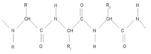
Figure 1 - Primary structure of keratin
Cysteine occurs regularly in keratin. These sulfur-containing amino-acid units cross-link with one another by forming disulfide bridges. The greater amount of cysteine, abundant in higher organisms, distinguishes keratin from elastins and collagens.
Because keratin's stability depends on the quantity of sulfur-containing amino acids, it can be divided according to their amount:
Soft keratin - keratin containing no more than 2% sulfur components.
Hard keratin - keratin containing up to 22% sulfur components.
Soft Keratin
It is easier to degrade. It occurs mainly in the lower layer of the epidermis and in the hair medulla. Its sulfur components are only weakly linked.
Hard Keratin
This type is highly resistant to chemical factors and is found primarily in nails, horns, and hair. Another notable feature is the pronounced, regular occurrence of serine. Like cysteine, this amino acid is responsible for hydrogen-bond formation and contributes to the secondary structure. Keratin is also characterized by elevated amounts of glycine and proline and reduced amounts of lysine, histidine, and methionine.
Proline deserves particular attention because it determines keratin's secondary structure (Schor and Krimm, 1961). It occurs in variable quantities of 8-12% and is responsible for keratin's right-handed coil. Table I summarizes the amino-acid composition of feathers.
Table I - Individual amino acids in keratin (Mukesh et al., 2012).
| Amino acid | Amount in feathers [g/kg protein] |
|---|---|
| Alanine | 56.6 |
| Arginine | 67.4 |
| Aspartate | 68.0 |
| Cysteine | 46.1 |
| Glutamate | 102.5 |
| Glycine | 76.6 |
| Histidine | 14.4 |
| Isoleucine | 49.2 |
| Leucine | 84.3 |
| Lysine | 22.3 |
| Methionine | 6.4 |
| Phenylalanine | 52.2 |
| Proline | 90.8 |
| Serine | 114.4 |
| Threonine | 48.9 |
| Tyrosine | 24.1 |
| Valine | 76.1 |
3. Secondary Structure
Keratin's secondary structure is determined primarily by its amino-acid composition. Four spatial conformations occur:
- alpha-keratin
- beta-keratin
- gamma-keratin
- amorphous keratin
Alpha-keratin is the most common. This conformation has a right-handed alpha-helical spatial arrangement and an average molar mass of about 60-80 kDa. One helical turn requires 64 amino-acid residues and has a length of 189 A (Schor and Krimm, 1961). It occurs most commonly in mammals.
Beta-keratin is arranged as a beta-pleated sheet. It is most abundant where the cuticle needs protection. Beta-keratins are difficult to extract from samples and occur most commonly in reptiles and birds.
Gamma-keratin is globular, has a high sulfur content, and has a lower molecular mass, typically around 15 kDa. All other keratin structures lacking regularity are classified as amorphous keratin.
4. Tertiary Structure
Keratin's tertiary structure consists of three right-handed alpha-helices forming one left-handed superhelix. Cross-linking through disulfide bridges also contributes to this structure.
Four such superhelices twisted together form a protofibril, the basic unit of microfibrils. Two central microfibrils surrounded by seven others form a macrofibril, which can be observed with a light microscope. The entire complex is stabilized by the previously mentioned disulfide bonds and by hydrogen bridges (Korniłłowicz-Kowalska and Bohacz, 2011). Figure 2, reproduced from the publication, summarizes the structural levels of keratin.
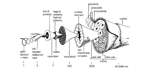
Figure 2 - Structural levels of keratin (Korniłłowicz-Kowalska and Bohacz, 2011).
I. Use of Keratin-Containing Waste Materials
Human consumption of chickens produces enormous quantities of waste feathers every day. Burning them is not only uneconomical but also fails to use the material's hidden potential fully. The United States alone produces approximately 815 million kg of feathers annually (Ichida and Krizova, 2001), while Poland produces 77,000 tonnes of broiler feathers per year (Korniłłowicz-Kowalska and Bohacz, 2011). Feathers account for about 5-7% of a chicken's weight. Thus, 50,000 poultry birds yield an average of approximately 2-3 tonnes of feathers (Dalev, 1994), enough to justify finding a more useful processing method.
3. Keratin as Feed
A feather hydrolysate produced by treating feathers with alkaline proteinase under alkaline conditions appears promising. Such a hydrolysate contains amino acids in a digestible form.
The first pilot study was carried out in 1971 (Elmayergi and Smith, 1971), demonstrating how feathers could be used to feed chickens and the nutritional value of feathers in their diet. Feather hydrolysate was fermented with Streptomyces fradiae, then fed to chickens. The fermented feather mixture produced greater weight gain than unfermented feathers; fermented feathers supplemented with essential amino acids produced a gain of 68.4 +/- 1.5 g, compared with 59.0 +/- 4.7 g for soy. However, the feed's nutritional value did not otherwise improve, probably because the chickens found the post-fermentation mixture unacceptable in the form obtained.
Another experiment fed young chickens equal amounts of different food for three weeks after three days of fasting. Soybeans served as the control, the second group received feather keratin meal, and the third received the same meal with keratinase from Bacillus licheniformis PWD-1. Daily weight gains were 66, 50, and 56 g respectively (Patent No. 5186961).
In a 2009 experimental study (Seo et al., 2009), pigs were fed for 11 weeks with feather meal prepared by degrading keratin under high pressure and boiling. A corn and soy mixture served as the control. Feed consumed per unit of weight gain for the control, 3% feather meal, and 6% feather meal respectively was 2.48, 2.41, and 2.45 in weeks 0-3; 2.81, 3.14, and 3.00 in weeks 3-8; 3.02, 3.16, and 3.24 in weeks 8-11; and 2.80, 2.95, and 2.93 in weeks 10-11. The results show a slight increase in weight when feed was enriched with feather meal. Keratin is also used in cosmetics, where it is added to hair-strengthening shampoos, skin creams, lubricating oils, and other creams.
II. Methods of Feather Hydrolysis
Keratin can be degraded effectively when its disulfide bridges are broken with reducing agents such as CuSO4, mercaptoacetate, iodoacetic acid, sodium sulfide, or sodium tetrathionate. Mass reduction of this kind is impractical industrially because of its high cost and because the intermediate would be difficult to process with microorganisms, since reducing agents are toxic to most of them. These agents are also poisonous, so the resulting material could not be used as feed.
1. Physical Degradation
Hydrothermal degradation uses high pressure and steam. Although suitable for breaking down feathers, it has not proved practical because of high energy costs and because amino acids such as methionine, lysine, and tryptophan are destroyed, while undesirable amino acids such as lysinoalanine and lanthionine form. These by-products reduce the nutritional value of the mixture.
2. Chemical Degradation
Chemical degradation generally hydrolyzes bonds at high or extreme pH. Acid hydrolysis using HCl at elevated temperatures, formerly used in producing food flavorings, can degrade keratin but is not very effective. The high acid concentrations required destroy important amino acids. The other method uses hydroxides in an alkaline environment, sometimes with elevated pressure or temperature. Alkalinity and pressure disrupt disulfide and peptide bonds, but this traditional method has not become widespread because it requires substantial energy and often destroys nutritionally valuable lysine, histidine, and methionine while producing lysinoalanine, lanthionine, and other compounds.
3. Enzymatic Degradation
Enzymatic degradation is a reasonable alternative because it requires lower temperatures and fewer chemicals. Most enzymes used in detergents are modified to work in alkaline environments at elevated temperatures.
Alcalase (National Center for Biotechnology Education, 2013) is a commercial enzyme produced by Bacillus licheniformis. It is a serine endopeptidase with very broad substrate specificity. Its optimum pH is 6.5-8.5, its optimum temperature is 45-65 C, and its maximum activity occurs at 60 C. Its converted price is approximately CZK 350 per 100 ml.
Savinase (National Center for Biotechnology Education, 2013) is a commercial enzyme produced by Bacillus species such as Bacillus lentus. It is also a broadly specific serine endopeptidase. Its optimum pH is 8-12, its optimum temperature is 20-60 C, and its maximum activity occurs at 55 C. Its converted price is approximately CZK 350 per 100 ml.
Esperase, most often produced by Bacillus species, is another enzyme that could replace keratinase. Enzymatic degradation alone is limited because disulfide bridges reinforce keratin so strongly that enzymes cannot degrade enough of it quickly without pretreatment.
4. Enzymatic-Chemical Method
In the first step, 500 g of feathers was boiled for 30 minutes with one liter of 0.3 M NaOH at 180 C. The temperature was then lowered and 1.25 g of alkaline proteinase was added. Bonds were hydrolyzed for two hours, after which the proteinase was inactivated by vigorous boiling. After drying, the 500 g of processed feathers yielded 19.0001 g of a gray powder with a density of 0.332 g/cm3, a salty taste, and a characteristic odor (Mukesh et al., 2012).
The mixture was analyzed and compared with feathers degraded in 6 M HCl for 24 hours at 110 C. The hydrolysate and HCl-treated feathers respectively contained, in g amino acid/kg feathers: alanine 55.6/58.9; arginine 70.1/67.7; cysteine 42.4/44.7; glutamate 102.8/101.5; glycine 76.9/76.5; histidine 13.7/14.1; isoleucine 51.5/49.1; valine 69.9/74.1; leucine 86.1/84.2; lysine 23.7/22.1; phenylalanine 50.5/52.1; proline 89.2/90.5; serine 112.5/114.2; threonine 44.5/48.7; and tyrosine 24.5/24.2.
The amino-acid composition remained stable, so the feathers did not lose nutritional value; the principal difference was consistency. Water was measured after three hours of drying at 105 C. Fiber was measured by extraction in 2% H2SO4 and 2% KOH at 100 C for 15 minutes. Fat was measured by ether extraction. Ash was weighed after burning the material at 550-600 C for three hours. Minerals were analyzed after digesting the starting material with HNO3 and HClO4, using argon emission spectrometry.
The dried hydrolysate and feathers respectively contained, in mg/g dry matter: water 51/494; crude protein 91/890; fiber 6.4/0; fat 13.2/14.3; ash 85.2/62.1; calcium 3.2/3.4; phosphorus 0.9/1.1; sodium 14.4/4.6; and chloride ions 20.7/7.6.
The main difference lies in the form in which the amino acids occur. Feather hydrolysate appears to be an interesting alternative poultry feed because poultry can digest it readily. A discrepancy was found when comparing the results: Dalev (1994) gives crude protein as 894.4 mg/g, whereas Kumar (2012) gives 91 mg/g. This discrepancy should be verified experimentally.
III. References
Avasn M., Aruna L., Ramakrishna R., Apta C. (2011) Degradation of feather and hair by Chrysosporium tropicum: A potent keratinophilic fungus. African Journal of Biotechnology, 10, pp. 3579-3584.
Burtt E., Ichida J. (1999) Occurence of feather degrading bacilli in the plumage of birds. The Auk, 116, pp. 364-372.
Coward-Kelly G., Chang V., Agbogbo F., Holtzapple H. (2006) Lime treatment of keratinous materials for the generation. Bioresource technology, 97, pp. 1337-1347.
Dalev P. (1994). Utilization of waste feathers from poultry slaughter for production of a protein concentrate. Bioresource technology, 45, pp. 265-267.
Deivasigamani B., Alagappan, M. (2008). Industrial application of keratinase and soluble proteins from feather keratins. Journal of enviromental biology, pp. 933-936.
Elmayergi H., Smith R. (1971). Influence of growth of Sreptomyces fradiae on pepsin-HC1 digestibility of feather meal, Journal of Microbiology, pp. 1067-1072.
Hill P. (2010). Some properties of keratin biomaterials: Kerateines. Biomaterials, pp. 585-593.
Ichida J., Krizova L. (2001). Bacterial inoculum enhances keratin degradation and biofilm formulation in poultry compost. Journal of microbiological methods, pp. 199-208.
Korniłłowicz-Kowalska T., Bohacz J. (2011). Biodegradation of keratin waste: Theory and practical aspects. Waste Management, 31, pp. 1689-1701.
Lin X. (1992). Purification and characterization of a keratinase from a feather degrading Bacillus licheniformis strain. Appl.Environ. Microbiol (58), pp. 3271-3275.
Maruthi A. (2011). Degradation of feather and hair by Chrysosporium tropicum. A potent keratinophilic fungus. African Journal of Biotechnology, 10, pp. 3579-3584.
Mukesh K., Lavanya P., Immaculate R., Balakumaran D., Kalaichelvan P. (2012) Production of Feather Protein Concentrate from Feathers by In vitro Enzymatic Treatment, its Biochemical Characterization and Antioxidant. Nature, Middle-East journal of Scientific research, 11, pp. 881-886.
Novoeznyme - Savinase [online]. Updated 16 January 2013, accessed 16 January 2013. Available at http://www.ncbe.reading.ac.uk/ncbe/materials/enzymes/savinase.html)
Onifade A., Al-Sane N., Al-Musallam A., Al Zarban S (1998) Review: Potentials for biotechnological applications of keratin-degrading microorganisms and their enzymes for nutritional improvement of feathers and other keratins as livestock feed resources. Bioresource technology, 66, pp. 1-11.
Patent No. 5186961: method and composition for maintainning animals on a keratin-containing diet, 1993-02-16.
Protease - novoenzyme Alcalase [online]. Updated 16 January 2013, accessed 16 January 2013. Available at http://www.ncbe.reading.ac.uk/ncbe/materials/enzymes/alcalase.html
Rogers G., Kemp J. (1972) Differentiation of Avian Keratinocytes. Characterization and relationships of the Keratin proteins of adult embrionic feathers and scales. Biochemistry, 11, pp. 969-975.
Seo S., Jung B., Lee M., Paik K. (2009) The Effect of Dietary Supplementation of Feather Meal on the Performance and Muscular Taurine Contents in Growing-finishing pigs, Asian-Aust. J. Anim. Sci., 22, pp. 1407-1413.
Sharma, Mu., Sharma Me., Rao V. (2011) In vitro biodegradation of keratin by dermatophytes and some soil keratinophiles. African Journal of Biochemistry Research, 5, pp. 1-6.
Schor R., Krimm S. (1961). Studies on the structure of feather keratin. Biophysical Journal, 1, pp. 489-515.
Wrześniewska-Tosik R., Janusz A. (2007) Biocomposites with a content of keratin from chicken feathers. fibres & textiles in Eastern Europe, 15, pp. 106-112.
Xing Z. (2011) Keratin Nanofibers as a Biomaterial. International Conference on Nanotechnology and Biosensors, pp. 120-124. Singapore.